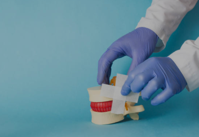
Both kyphoplasty and vertebroplasty allow physicians to cement a fractured bone in place. Patients often experience significant pain relief as well as increased mobility. These procedures are performed under general anesthesia or conscious sedation. During the process, an interventional radiologist inserts a hollow needle into the damaged bone. They may insert a balloon first and then fill the space with a bone cement mixture to reduce unwanted kyphosis, strengthen the spine, and relieve pain.
Vertebroplasty
Before your procedure, follow your doctor’s instructions about taking medications and preparing for surgery. You may need to avoid eating or drinking anything other than small sips of water for several hours before your procedure. You should also wear loose, comfortable clothing and leave jewelry at home. If you take blood pressure medicine, ask your doctor if it is safe to take it on the day of your procedure. You will be put under general anesthesia. A trocar, a hollow needle or tube, is introduced through your skin and into the fractured vertebra under X-ray guidance. Then, orthopedic cement is injected into the spine, quickly hardening and stabilizing the vertebra. During kyphoplasty, physicians insert a balloon through the hollow needle and then inflate it inside the collapsed vertebra to elevate and restore some of its height. Once the balloon is removed, bone cement is injected into the created space. The cement will help the vertebral body stay upright and relieve pain.
Kyphoplasty
Kyphoplasty Jacksonville FL is an outpatient procedure that strengthens weakened spines and repairs fractured vertebrae. It can also help improve pain and posture. It is usually recommended if other treatments, such as back braces, steroid injections and pain medications, haven’t worked. In a kyphoplasty, the surgeon first numbs the skin with a local anesthetic. Next, a hollow trocar needle is inserted into the collapsed vertebrae body using imaging guidance. A balloon-like device called a balloon tamp is then inserted through the trocar and inflated, creating space for the cement to be injected. The balloon is then removed. The doctor fills the space in the vertebrae with a bone cement mixture and then re-inflates the balloon to create more space. It allows them to inject more cement if necessary. The operation takes around an hour per vertebra. Patients may feel sore or have a slight backache where the trocar was inserted, but this should improve within a few days.
Post-Procedure Care
An intravenous line is inserted into a vein in your arm before the treatment to administer painkillers and sedatives. The doctor will also place a needle in the back and, using X-ray guidance, insert a balloon into the bone. The balloon inflates, creating a space and helping the vertebrae regain shape. The broken vertebrae are subsequently filled with medical-grade bone cement. The procedure is usually finished within 60 minutes.
After the procedure, you’ll be taken to a recovery room. The region where the incision was made is likely to be sore, and your doctor might advise applying ice to it for one to two hours each day.
Your doctor would advise painkillers and a physical therapy regimen to improve your spinal muscles. You may need to prevent further back problems, such as avoiding strenuous activities and taking calcium supplements to avoid osteoporosis. It is especially important if you have a history of vascular conditions like a stroke or heart disease that increases your risk for bone fractures.
Recovery
As with any procedure, recovery from Kyphoplasty/Vertebroplasty will take time. However, since it is minimally invasive, patients typically experience rapid pain relief and go home the same day, making it an outpatient procedure. The procedure involves having you lay on your stomach while a medical professional inserts a trocar—a hollow needle—through your skin and into the collapsed vertebra. It is done using a type of X-ray called fluoroscopy to ensure the hand is correctly placed. Once positioned, bone cement is injected under visual volume and pressure controls into the space created by the trocar. Once the cement has been injected, it can harden; the needles are removed. The small incision site is then closed with skin glue or sterile strips. Once the surgery is over, you will be transferred to a recovery room for one or two hours. The doctor will monitor your condition until the anesthesia wears off.


